INTRODUCTION
Certain breeds (e.g. Siberian Husky), old dogs and
not neutered males are more prone to perianal
(circum-anal) gland tumours. The majority of tumours are
benign and are called perianal (circum-anal) adenomas.
The malignant ones are called adenocarcinomas. They
can't be differentiated by visual inspection. A
histopathology of the excised tumour is needed to check
whether it is cancerous.
 TREATMENT - 3 OPTIONS TREATMENT - 3 OPTIONS
1. Surgical excision.
Some vets don't
perform this surgery as there may be worries of the
wound not being able to heal and close. The client may
get very angry as the dog will keep licking the open
wound. Old dogs may die under general anaesthesiaC on the
operating table leading to highly emotional scenes and
potential litigation.
These are two main reasons why some vets don't want to
operate. The owner
may need to seek a vet who will operate as the dog
licks the infected tumours to relieve its pain. Blood
dripping from the backside can be quite inconvenient to
the owner. Owners may need to be proactive in seeking
early surgical removal.
2. Neutering.
Perianal gland tumours are most common in male dogs that
are not neutered. It is rarer in female dogs. In this
case study in 2010, a female spayed Siberian Husky had one such
tumour.
3. Hormone treatment.
Tardak is an anti-androgenic hormone. Injections may be
effective but need to be given regularly when tumours
occur.
Tardak is for use in male dogs and cats in the following
indications:
3.1 The treatment of hypersexuality (humping, wanting to
stray).
3.2 The relief of prostatic hypertrophy whether benign,
carcinomatous or when due to chronic inflammatory
processes. In inflammation, antibiotics and
anti-inflammatory drugs are used too.
3.3 For the treatment of
circum-anal (perianal) gland tumours.
3.4 For the treatment of certain forms of
aggressiveness, nervousness, epileptiform seizures and
corticoid-resistant pruritus (developing into dermatoses
and accompanied by alopecia).
4. Chemotherapy and radiation for cancerous types. This
is not normally available for Singapore dogs.
|
A 2010
CASE STUDY
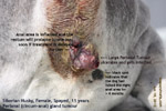
Female spayed Siberian Husky, 11 years old suffered from
blood dripping from her circum-anal tumour for over 4
months. The tumour ulcerates and become infected. If you see
the black spot below and to the right of the anus, there
were early signs of perianal gland irritation, as the
black spot is due to continual licking over several
weeks.
CHALLENGES
1. High anaesthetic risk as in all old dogs. The owner
did not wish to have a blood test. The vet must inform
the owner of the need for such a test to screen the
health of the dog before anaesthesia and surgery.
2. Large tumour over 3 cm x 3 cm very close to the anus.
That meant a large wound after removal of the tumour and
difficulty in achieving normal closure.
3. The dog passes soft stools during surgery, resulting in
possible contamination during surgery and after.
4. If the
dog rubs her backside on the floor after surgery, the
stitches may break down. A large e-collar prevents
licking of the wound.
PLAN AHEAD
The owner's vet who did not want to
operate. The dog had a full grooming esp. of the tail area,
ensuring that there were no maggot wounds.
I prescribed oral Baytril
antibiotics for 6 days.
PRE-ANAESTHESIA
On the 6th day, the wound was not infected. The dog's
rectal temperature could not be taken as the dog
struggled and leaked out urine whenever her tail was to
be held up for the insertion of the thermometer into the
rectum. She was in great pain in the anal area and tried
to bite to defend herself. More urine leaked out as she
struggled. I got her muzzled for the IV drip and the
injection via the IV catheter of Domitor 0.15 ml.
ANAESTHESIA
The dog was shaved. Isoflurane gas was given by mask. I intubated the dog and
gave isoflurane at 1-2% ensured
surgical anaesthesia. I got a towel to cover the
metallic operating table to prevent electrical shock
when I used electro-surgery to excise the
circum-anal
tumour. A swab with saline was placed on the indifferent
plate and the dog's belly for the conduct of electricity
during electro-incision and cautery.
SURGERY, JUNE 9, 2010
FROM 2.17 PM - 3.15PM
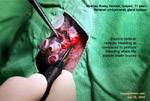
1. I used electro-incision to cut off the tumour. The
dorsal part has a dark red mass of 0.5 cm x 0.5 cm. The
main mass was hard, nodular and ulcerated. It was 2.5 cm
x 2.5 cm. The shorter the surgery, the better the
chances of survival.
2. A small artery at the ventro-lateral area nearer and
below the anus spurted out red blood. I ligated 3 times.
Coagulated the bleeding point. Finally, there was not
much bleeding.
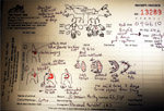
3. As the elliptical gap was large and under high
tension, I had to reduce the tension in order to enable
good wound healing. I extended the skin incision to the
left at the dorsal and ventral edges above and below the
anus respectively (see illustration). The wound was
stitched with 3/0 sutures.
 |
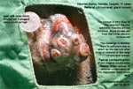 |
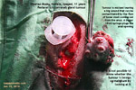
The anus must be plugged to
prevent stools from contaminating
the surgical wound with bacteria.
A 3-ml syringe was initially used.
|
|
|
|
|
 |
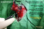 |
Electro-incision is very useful
when the tumour is one discrete
lump. No bleeding at all. The
profuse bleeding is due to a small
artery on the lower left shooting
out blood. It was ligated.
|
|
|
|
 |
Copious soft yellow stools leaked
out only during anaesthesia. I
switched to a 10-ml syringe but
still the stools flowed. In
retrospect, a spasmogesic
injection IV should be given in
this case. Anaesthesia was very
low. This could be deepened but
this old dog might just die.
|
|
|
|
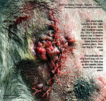 |
Note the loose reddish tissues
outside the anus. This indicated a
partial rectal prolapse due to
continual irritation of the rectum
by loose stools over the years. |
| |
|
|
I illustrate
the surgical techniques used for my record and
review. The owner was given the original copy.
This surgery can result in a big contaminated
wound and lots of cursing by the unenlightened
owner if the stitches break down and if the owner
does not do proper nursing consequentially.
|
 |
4. During closing, this dog kept passing out the loose
stools despite her rectum being plugged by a 3-ml
syringe. The stools would fall onto the surgical wound
and this would be terrible. The wound would be
contaminated and would not heal. "Get me a
10-ml
syringe," I asked my assistant. The 10-ml
syringe plugged the rectum. Still the
stools flowed out from the side. I had to be careful to
wipe the stools away while I quickly stitched up.
This was be possibly due to
the minimal isoflurane gas being used and therefore the
dog's defaecation reflex was present. The dog's tongue
was not a healthy pink and sometimes it turned cyanotic.
Since the owner did not want a health screening blood
test, it would be difficult to know if this dog had
anaemia, hypercalcaemia, kidney or liver disorders.
POST SURGERY
The dog woke up within 10 minutes as if she had a good
nap of over 30 minutes.
I gave 2.5 ml of anti-spasmogesic IV to prevent more
loose stools coming out to contaminate the wound. She
wore an e-collar. She was the nervous type. The
owners visited her
in the evening. "Was the lady owner able to check out
the surgical area?" I asked Mr Saw, my assistant. Every
time we tried to lift the tail, this dog would panic and
void a large volume of yellow urine. I phoned the owner
to take the dog home as this seemed to be in the
interest of the dog.
HISTOPATHOLOGY
The vet must inform the owner that there is histopathology
to verify whether the tumour is cancerous or benign. In
this case, the owner did not want this test.
CONCLUSION
Neutering and surgical excision are recommended. Tumours
are best removed when they are very small. I advise two
anti-androgenic injections post surgery at 2-weekly
intervals. Neutering your male dogs when they are young
and/or weekly examination of the anal area will ensure
that your dog live longer. This case has a happy ending. It is best that the
owner gets the tumours excised when they are very small
in size.
UPDATE ON JUN 10, 2010
1. The owner visited her yesterday evening after the
surgery. The dog permitted only the lady owner to lift
his tail to look at the surgical site. I suggested that
the dog go home.
2. The lady arranged for the dog transport man to bring
the dog home. She came to the Surgery at 5 pm on Jun 10,
2010 to wait for the transport man.
3. Although she did not pay for the blood test, I showed
her the results. The blood urea was high. It was 14.4 (normal =
4.2 to 6.3). Creatinine was within normal range
at 117 (89 -177).
Total White Cell
Count was
high at 18.2 (normal = 1 - 17)
despite 6 days of 2.5 tablets of Baytril
50mg for this 25 kg
weight, before the
blood test. Neutrophils 83.5% (normal
60-70%), Lymphocytes 6.7%, Monocytes 9.3%,
Eosinophils 0.06% and Basophils 0.55%. I
considered this result not to be a health
threat.
4. I advised the owner that continuous passage of loose
stools will result in rectal prolapse. She
confirmed that she saw some rectal tissues coming out of
the anus. It is possible that the dog may not tolerate
dry dog food or could be allergic to the components of
ordinary dry dog food. Passing loose stools over several
years may result in rectal prolapse or intusussception.
5. Unusual behaviour of this older dog. Whenever she is
restrained, she would "wet her pants". A lot of urine
would shoot out. It seems that she withholds urine as
this incident happens more than 4 times during restraint
before and after surgery. This could be
a behaviour of excitation urination.
6. Home nursing. A small spray bottle filled with
warm water. Spray onto the surgical wound to remove
faeces or blood. Gently touch with tissue paper.
Painkillers tolfedine to be given for next 4 days.
E-collar to be worn. This dog was happier at home as
she
seemed to be frightened of veterinary staff and vets.
7. I hope all will be well with this old dog and that
the tumour will not recur. A blood health screening
in older dogs will always be good for the dog and the
owner as diseases shown can be treated or prevented.
UPDATE ON JUN 10, 2010
The lady owner phoned me. "The stitches have broken
down but the dog will not allow me to lift the tail to
see the wound."
"Don't worry," I said. "The tolfedine pain-killers would
work soon. Did the dog pass stools?"
"Yes, one small brown bit. Solid stool."
"That's good," I said. The dog was off dry food for the
past 48 hours. My spasmogesic injection IV had slowed
down intestinal peristalsis permitting the stool to be
well formed. Did the
stitch break down due to the dog licking the wound? I
forgot to ask whether the dog was wearing an e-collar.
No further news from the owner.
|
 TOA
PAYOH VETS
 TOA
PAYOH VETS